[ad_1]

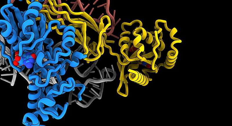
Using electron microscopy, scientists have managed to create a 3D model of a part of a human cell, the ribosome, which is no more than 30 nanometers in diameter. Credit: Eva Kummer
Human cells contain ribosomes, a complex machine that makes proteins for the rest of the body. Researchers have now come closer to understanding how the ribosome works.
“It’s amazing that we can see the atomic details of ribosomes. Because they are small – about 20-30 nanometers,” says Associate Professor Eva Kummer from the Novo Nordisk Foundation Center for Protein Research. A new study Published in Nature Communications.
A ribosome is a part of a human cell that contains ribosomal RNA and ribosomal proteins. It is like a factory that makes proteins by following the set of instructions contained in the genes.
Ribosomes are found floating in the cell cytosol, cellular organelles such as mitochondria or the protoplasm of bacteria.
to use Electron MicroscopyKummer and his colleagues Giang Nguyen and Christina Ritter have managed to create a 3D model of a part of the human cell, the ribosome, which is no more than 30 nanometers in diameter.
More specifically, they have taken snapshots of how the ribosome is made.
“It is important to understand how the ribosome is formed and how it works, because it is the only particle in the cell that makes proteins in humans and all other organisms. And without proteins, life would cease to exist.” The waist is called.
Proteins are the basic building blocks of the human body. Your heart, lungs, brain, and basically your entire body are made of proteins made by ribosomes.
“From the outside, the human body looks very simple, but then consider the fact that every part of the body is made up of millions of molecules, which are extremely complex, and that they all know what to do – that’s enough. It’s breathtaking.” waist
Fold, assemble, and move to the right place
Before The ribosome Before proteins can begin to produce, they first need to be assembled from more than 80 different components.
Kummer and his colleagues obtained 3D models of three different stages of ribosome assembly. “It’s a complex particle with many different parts—many protein and RNA components—that must be folded, assembled, and transported to the right place. It doesn’t all happen at once. Ribosome assembly is a gradual process. is a process that involves several steps,” she explains.
Of the three steps, the 3D model that describes the earliest time point in assembly is the most interesting, according to Kummer, because no one has been able to describe it before.
“At this stage, we can tell, for example, that a specific protein called GTPBP10 is eager to interact with a so-called RNA component that forms a long helix,” says Kummer. “In fact, towards the bottom of this helix is the catalytic center of the ribosome, where proteins are made. That’s why it’s so important that the helix is folded and positioned correctly.”
To achieve this, GTPBP10 grasps the helix and holds it in the correct position. Protein synthesis.
This is just one of many steps in ribosome assembly that new research has shed light on—insights that could pave the way for learning more about various diseases.
“Defects in ribosome assembly severely reduce the ability of our cells to make proteins. For example, it is the proteins that convert the energy we get from food into energy coins that the body uses in all cellular types. can use to run the process.
“Now, if the mitochondrial ribosomes don’t work, our body can’t produce more energy and this leads to diseases like neurodegenerative disorders and heart disease,” says Kummer. “The first step is to understand how things work.” They work. Only then can you try to change them.”
More information:
Thu Giang Nguyen et al., Structural insights into the role of GTPBP10 in mitoribosome RNA maturation, Nature Communications (2023). DOI: 10.1038/s41467-023-43599-z
Provided by
University of Copenhagen
Reference: Researchers create 3D model of ribosome and visualize how it forms (2024, February 23) Retrieved February 24, 2024 from https://phys.org/news/2024-02-3d-ribosome-visualize.html Obtained
This document is subject to copyright. No part may be reproduced without written permission, except for any fair dealing for the purpose of private study or research. The content is provided for informational purposes only.
[ad_2]


